Gharbi Classification : Classification of hydatid cysts according to Gharbi and ... / Fluid collection with a split wall fluid collection with septa heterogenous echo patterns.
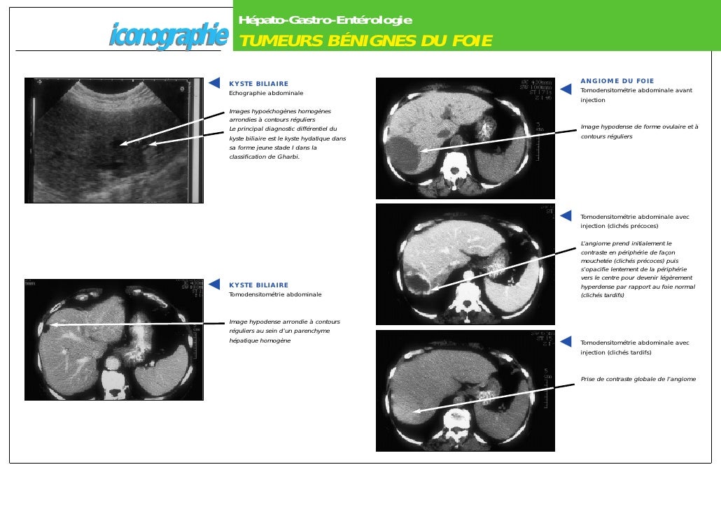
Gharbi Classification : Classification of hydatid cysts according to Gharbi and ... / Fluid collection with a split wall fluid collection with septa heterogenous echo patterns.. Then, the diagnosis of pleural primitive hydatid cyst type ii of gharbi classification was retrospectively made. Ultrasound is also helpful during treatment monitoring while on medical treatment and also for postoperative follow up (6, 7). The initial classification by gharbi et al and the who informal working group on. There are several classification schemes for liver hydatid cysts based on their ultrasound appearances; With muscle hydatidosis were classified according to the gharbi classification (table 1).
The liver segments were grouped as near to the hilum (segments i, iii, ivb, v, and vi) and remotely distant (segment ii, iva, vii. Gharbi classification → in 1981 hassen gharbi classified liver hcs into five types based on ultrasound findings → type i: Surgical treatment was used for all cysts that were larger than 15 cm, those that were complicated, and those that were not. Classification systems based on the us appearance of the cyst have been developed for ce and ae, which may provide important information to the physician managing the case. In order to assess the relation of the cyst with the biliary tract, the relevant ducts were visualized following the irrigation of the cavity via isotonic.
Liver abscesses and hydatid disease from image.slidesharecdn.com Then, the diagnosis of pleural primitive hydatid cyst type ii of gharbi classification was retrospectively made. In his classification, he considers five types according the natural evolution of the parasite. Gharbi classification → in 1981 hassen gharbi classified liver hcs into five types based on ultrasound findings → type i: In order to assess the relation of the cyst with the biliary tract, the relevant ducts were visualized following the irrigation of the cavity via isotonic. In 1981 professor gharbi et al. The initial classification by gharbi et al and the world health organization. Ultrasound examination of the hydatic liver. Proposed a us classification of the hydatid cysts 10.
Myriam gharbi honorary research associate, joseph h drysdale student, hannah lishman research assistant, rosalind goudie research assistant, mariam molokhia clinical reader, alan p johnson.
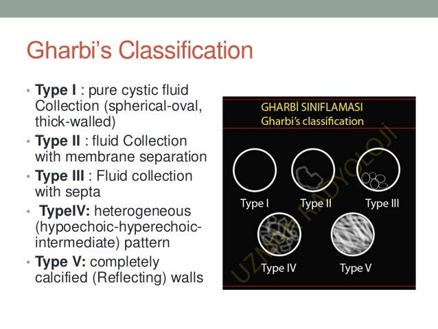
There are several classification schemes for liver hydatid cysts based on their ultrasound appearance; Cyst's type according to gharbi classification. In 1981 professor gharbi et al. Abdominal u&s and ct were used as the radiological diagnostic tools in all patients, according to the gharbi classification in s&u, six of the cysts were type 2, and two of the cysts were type 1. On the other hand gharbi classification also divides in 5 types: Then, the diagnosis of pleural primitive hydatid cyst type ii of gharbi classification was retrospectively made. Type i cysts consist of pure fluid; The initial classification by gharbi et al and the who informal working group on. Several classifications of chd exist. With muscle hydatidosis were classified according to the gharbi classification (table 1). Ultrasound is also helpful during treatment monitoring while on medical treatment and also for postoperative follow up (6, 7). The gharbi classification was used to stage the cysts 8. Type ii has a fluid collection with a split wall;
Surgical treatment was used for all cysts that were larger than 15 cm, those that were complicated, and those that were not. Proposed a us classification of the hydatid cysts 10. Abdominal u&s and ct were used as the radiological diagnostic tools in all patients, according to the gharbi classification in s&u, six of the cysts were type 2, and two of the cysts were type 1. In order to assess the relation of the cyst with the biliary tract, the relevant ducts were visualized following the irrigation of the cavity via isotonic. On the other hand gharbi classification also divides in 5 types:
Iconographies from image.slidesharecdn.com Proposed a us classification of the hydatid cysts 10. Myriam gharbi honorary research associate, joseph h drysdale student, hannah lishman research assistant, rosalind goudie research assistant, mariam molokhia clinical reader, alan p johnson. Correspondence between gharbi and who classification of liver hydatid cysts. Classification of hydatid cysts based on ultrasonographic findings. Type i cysts consist of pure fluid; Cysts were classified according to the gharbi classification in 15 studies (27.8%) and according to the the who has classified hepatic ce based on ultrasonographic features of the cyst 6. On the other hand gharbi classification also divides in 5 types: 12 those with gharbi type iii cysts were treated on an outpatient basis.
In order to assess the relation of the cyst with the biliary tract, the relevant ducts were visualized following the irrigation of the cavity via isotonic.
Classification systems based on the us appearance of the cyst have been developed for ce and ae, which may provide important information to the physician managing the case. Gharbi classification → in 1981 hassen gharbi classified liver hcs into five types based on ultrasound findings → type i: Gharbi classification pure fluid collection. Classification of hydatid cysts based on ultrasonographic findings. Pure fluid collection → type ii: Proposed a us classification of the hydatid cysts 10. (1) hypoechoic appearance (2) hyperechoic solid pattern without back. Abdominal u&s and ct were used as the radiological diagnostic tools in all patients, according to the gharbi classification in s&u, six of the cysts were type 2, and two of the cysts were type 1. The liver segments were grouped as near to the hilum (segments i, iii, ivb, v, and vi) and remotely distant (segment ii, iva, vii. Type iii cysts contain daughter cysts (with or without. In 1981 professor gharbi et al. Who introduced a standardized classification of ultrasonography images of cystic echinococcosis, to. Human body anatomy ct scan cysts pelvis abdomen abdominal.
The initial classification by gharbi et al and the world health organization. Type iii cysts contain daughter cysts (with or without. Classification systems based on the us appearance of the cyst have been developed for ce and ae, which may provide important information to the physician managing the case. Human body anatomy ct scan cysts pelvis abdomen abdominal. Ultrasound examination of the hydatic liver.
Characteristics of cysts: size, location, sonographic ... from www.researchgate.net Ultrasound is also helpful during treatment monitoring while on medical treatment and also for postoperative follow up (6, 7). Type i cysts consist of pure fluid; The gharbi classification system was used to stage the hydatid disease 5. Classification of hydatid cysts based on ultrasonographic findings. With muscle hydatidosis were classified according to the gharbi classification (table 1). International classification of ultrasound images in cystic echinococcosis for application in clinical and field epidemiological settings // acta trop. 12 those with gharbi type iii cysts were treated on an outpatient basis. Who introduced a standardized classification of ultrasonography images of cystic echinococcosis, to.
In order to assess the relation of the cyst with the biliary tract, the relevant ducts were visualized following the irrigation of the cavity via isotonic.
Ultrasound examination of the hydatic liver. In his classification, he considers five types according the natural evolution of the parasite. Classification systems based on the us appearance of the cyst have been developed for ce and ae, which may provide important information to the physician managing the case. Abdominal u&s and ct were used as the radiological diagnostic tools in all patients, according to the gharbi classification in s&u, six of the cysts were type 2, and two of the cysts were type 1. The initial classification by gharbi et al and the world health organization. The gharbi classification was used to stage the cysts 8. There are several classification schemes for liver hydatid cysts based on their ultrasound appearance; Cyst's type according to gharbi classification. There are several classification schemes for liver hydatid cysts based on their ultrasound appearances; Type iii cysts contain daughter cysts (with or without. Hence, ultrasound is the primary imaging modality. The patient was put on albendazole for 6 months. Several classifications of chd exist.
The initial classification by gharbi et al and the who informal working group on gharbi. Fluid collection with a split wall.


0 Komentar